活細胞影像觀察—研究界的直播主:Chromobody


![]()
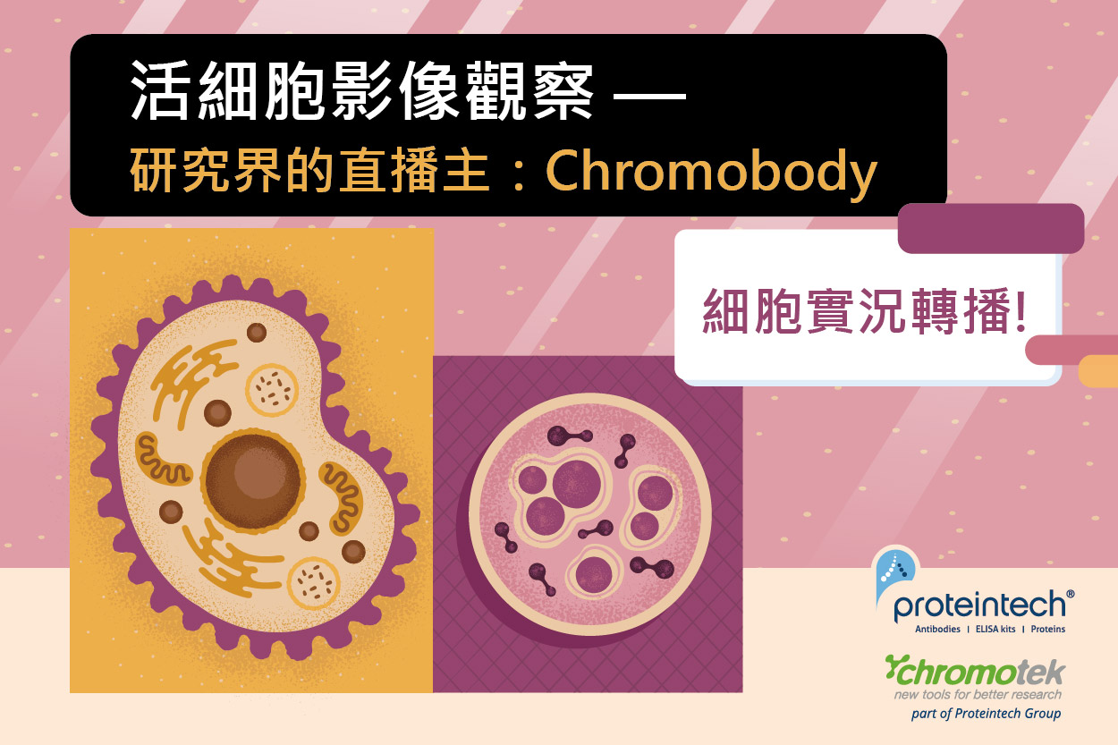
活細胞影像觀察— 研究界的直播主:Chromobody

▲(圖一)
應用無極限! 只有奈米抗體能夠超越自己!奈米抗體內標化:基於奈米抗體為單體、重組表達的特點,與其輕盈的體積,Chromotek將奈米抗體的序列編碼與螢光蛋白的序列融合,放置於表達的質體 (plasmid) 中,打造出可轉染至細胞,直接於活細胞中表現與螢光蛋白偶聯的奈米抗體的Chromobody (圖一),實現直接螢光標定活細胞中的內源蛋白質,目標內源蛋白的一舉一動實況轉播(圖二)!
 |
|
 |
Time lapse video of Actin-Chromobodies expressed in HeLa cells, which have been exposed to Cytochalasin D treatment for 1 hour and then recovered for 4 hours. |
 |
Cell Cycle-Chromobody expressed in HeLa cells. The time lapse video shows cell division. The signal of TagRFP has been imaged |
▲(圖二)
奈米抗體的靈巧體積讓活細胞感覺好自在
Chromobody經過多重驗證,不僅可有效、專一地與目標蛋白結合,更不會干擾其生物學功能,Melak et al. 2017的文獻指出與多種標定Actin的方法(圖三)相比,Actin-Chromody是對Actin的干擾最小的標定方式 (Table 1)。

▲(圖三)

▲Table 1: Overview of stte-of-the-art technologies/probes to visualize actin filaments:
Melak M, Plessner M, Grosse R. Actin visualization at a glance. J Cell Sci. 2017 Feb 1;130(3):525-530. doi: 10.1242/jcs.189068. Epub 2017 Jan 12. Erratum in: J Cell Sci. 2017 May 1;130(9):1688. PMID: 28082420.
使用Chromodoby,不用導致:
- 細胞生存異常、遷移
- 遺傳物質改變、交錯
- 影響細胞功能
- 在過度表達螢光融合蛋白時顯示細胞毒性效應
成就高解析美圖美景,不用美圖修修
如同螢光奈米二抗的最大優勢:其靈巧的體積等同於減少抗原表位和訊號標記物之間的距離 (圖四),是高解析率顯微鏡的理想探針,例如: STED (STimulated Emission Depletion (縮寫STED))、STORM (Stochastic Optical Rwjeconstruction Microscopy , 隨機光學重建顯微鏡系統) 等系統。用於活細胞動態攝影則大大降低影像模糊、動態殘影等影響後續分析的問題。

▲(圖四)
更多高解析影像系統的介紹,可點擊先前螢光奈米二抗的文章查看 ↓
奈米螢光二抗- 高解析度成像的次世代二級奈米抗體 (abrealbiotech.com)
Chromotek目前已推出數款Chromobody,適合與其他細胞標的物搭配,趕快來看看,活體影像直播開起來!
 |
用於視化活細胞骨骼和監測其動力學。Actin-Chromody不僅可用在哺乳動物細胞,而且在特殊物種,如斑馬魚和植物的細胞和組織中,對microfilaments進行非侵入性標記。 |
 |
用於即時觀察細胞大小、細胞核形態和線粒體。此外,可用於高含量分析(HCA)。 |
 |
追蹤活細胞細胞骨骼的動態。與主要存在於間質細胞中的Vimentin intermediate filaments結合。 |
 |
用於標定PCNA,是一種環繞 DNA 的同質環狀蛋白質,參與細胞週期中DNA各種的狀態變換。PCNA-Chromobody常用於探討癌症藥物等化合物對細胞週期和毒性的影響。 |
 |
PARP1是細胞核中最豐富的蛋白質之一,參與許多細胞過程,如DNA修復、轉錄調節和色素結構調製。PARP1-Chromobody可用於研究微輻射後的DNA損傷。 |
 |
Dnmt1 1 是真核細胞中的主要 DNA 甲基轉移酶,用以保持全基因組的甲基化模式,在發育過程中,控制基因表達和基因組穩定的表觀遺傳機制。 |
| Product | Plasmids (Product citations in parentheses) |
| Actin-Chromobody® | TagGFP2 (15), TagRFP (5) |
| Nuclear Actin-Chromobody® | TagGFP2 |
| Cell Cycle-Chromobody® | TagRFP (11) |
| Dnmt1-Chromobody® | TagGFP2, TagRFP (1) |
| Histone-Chromobody® | eGFP (1) |
| Lamin-Chromobody® | TagGFP2 (5) |
| PARP1-Chromobody® | TagGFP2, TagRFP (2) |
| Vimentin-Chromobody® | TagGFP (2) |
多篇重量級文獻真實推薦
Actin-Chromobody
The Actin-Family Protein Arp4 Is a Novel Suppressor for the Formation and Functions of Nuclear F-Actin. (Yamazaki et al. Cell 2020) https://www.ncbi.nlm.nih.gov/pubmed/32204557
Characterization of 3D Printed Stretching Devices for Imaging Force Transmission in Live-Cells. (Mayer et al. Cell Mol Bioeng 2019) https://www.ncbi.nlm.nih.gov/pubmed/31719915
Actin chromobody imaging reveals sub-organellar actin dynamics. (Schiavon et al. bioRxiv 2019) https://www.biorxiv.org/content/10.1101/639278v3
Formin-2 drives polymerisation of actin filaments enabling segregation of apicoplasts and cytokinesis in Plasmodium falciparum. (Stortz et al. eLife 2019) https://www.ncbi.nlm.nih.gov/pubmed/31322501
Indirect visualization of endogenous nuclear actin by correlative light and electron microscopy (CLEM) using an actin‑directed chromobody. (Abdellatif et al. Histochem Cell Biol 2019) https://www.ncbi.nlm.nih.gov/pubmed/31154480
A Strategy to Optimize the Generation of Stable Chromobody Cell Lines for Visualization and Quantification of Endogenous Proteins in Living Cells. (Keller et al. Antibodies (Basel) 2019) https://www.ncbi.nlm.nih.gov/pubmed/31544816
A transient pool of nuclear F-actin at mitotic exit controls chromatin organization. (Baarlink et al. Nat Cell Biol 2017) https://www.ncbi.nlm.nih.gov/pubmed/29131140
Toxoplasma gondii F-actin forms an extensive filamentous network required for material exchange and parasite maturation. (Periz et al. Elife 2017) https://www.ncbi.nlm.nih.gov/pubmed/28322189
Actin visualization at a glance. (Melak et al. J Cell Sci 2017) https://www.ncbi.nlm.nih.gov/pubmed/28082420
Coordinate-targeted fluorescence nanoscopy with multiple off states. (Danzl et al. Nature Photonics 2016) https://www.nature.com/articles/nphoton.2015.266
Nuclear F-actin formation and reorganization upon cell spreading. (Plessner et al. J Biol Chem 2015) https://www.ncbi.nlm.nih.gov/pubmed/25759381
Live imaging of endogenous protein dynamics in zebrafish using chromobodies. (Panza et al. Development 2015) https://www.ncbi.nlm.nih.gov/pubmed/25968318
Fluorescent labelling of the actin cytoskeleton in plants using a cameloid antibody. (Rocchetti et al. Plant Methods 2014) https://www.ncbi.nlm.nih.gov/pubmed/24872838
Cell Cycle-Chromobody
The dynamic equilibrium of nascent and parental MCMs safeguards replicating genomes. (Sedlackova et al. bioRxiv 2019) https://www.biorxiv.org/content/10.1101/828954v1
A Multiplexed High-Content Screening Approach Using the Chromobody Technology to Identify Cell Cycle Modulators in Living Cells. (Schorpp et al. J Biomol Screen 2016) https://www.ncbi.nlm.nih.gov/pubmed/27044685
Nuclear actin modulates cell motility via transcriptional regulation of adhesive and cytoskeletal genes. (Sharili et al. Sci Rep 2016) https://www.ncbi.nlm.nih.gov/pubmed/27650314
Live imaging of endogenous protein dynamics in zebrafish using chromobodies. (Panza et al. Development 2015) https://www.ncbi.nlm.nih.gov/pubmed/25968318
Live cell imaging at the Munich ion microbeam SNAKE – a status report. (Drexler et al. Radiation Oncology 2015) https://www.ncbi.nlm.nih.gov/pubmed/25880907
Assessing kinetics from fixed cells reveals activation of the mitotic entry network at the S/G2 transition. (Akopyan et al. Mol Cell 2014) https://www.ncbi.nlm.nih.gov/pubmed/24582498
Quantitative live imaging of endogenous DNA replication in Mammalian cells. (Burgess et al. PLoS One 2012) https://www.ncbi.nlm.nih.gov/pubmed/23029203
Dnmt1-Chromobody
Long non-coding RNA PARTICLE bridges histone and DNA methylation. (O'Leary et al. Sci Rep 2017) https://www.ncbi.nlm.nih.gov/pubmed/28496150
Histone-Chromobody
Under the Microscope: Single-Domain Antibodies for Live-Cell Imaging and Super-Resolution Microscopy. (Traenkle & Rothbauer. Front Immunol 2017) https://www.ncbi.nlm.nih.gov/pubmed/28883823
Lamin-Chromobody
A Strategy to Optimize the Generation of Stable Chromobody Cell Lines for Visualization and Quantification of Endogenous Proteins in Living Cells. (Keller et al. Antibodies (Basel) 2019) https://www.ncbi.nlm.nih.gov/pubmed/31544816
Characterization of 3D Printed Stretching Devices for Imaging Force Transmission in Live-Cells. (Mayer et al. Cell Mol Bioeng 2019) https://www.ncbi.nlm.nih.gov/pubmed/31719915
A transient pool of nuclear F-actin at mitotic exit controls chromatin organization. (Baarlink et al. Nat Cell Biol 2017) https://www.ncbi.nlm.nih.gov/pubmed/29131140
Case study on live cell apoptosis-assay using lamin-chromobody cell-lines for high-content analysis. (Zolghadr et al. Methods Mol Biol 2012) https://www.ncbi.nlm.nih.gov/pubmed/22886277
Novel antibody derivatives for proteome and high-content analysis. (Schmidthals et al. Anal Bioanal Chem 2010) https://www.ncbi.nlm.nih.gov/pubmed/20372881
Targeting and tracing antigens in live cells with fluorescent nanobodies. (Rothbauer et al. Nat Methods 2006) https://www.ncbi.nlm.nih.gov/pubmed/17060912
PARP1-Chromobody
A New Nanobody-Based Biosensor to Study Endogenous PARP1 In Vitro and in Live Human Cells. (Buchfellner et al. PLoS One 2016) https://www.ncbi.nlm.nih.gov/pubmed/26950694
Vimentin-Chromobody
Real-time analysis of epithelial-mesenchymal transition using fluorescent single-domain antibodies. (Maier et al. Sci Rep 2015) https://www.ncbi.nlm.nih.gov/pubmed/26292717
Visualizing Epithelial-Mesenchymal Transition Using the Chromobody Technology. (Maier et al. Cancer Res 2016) https://www.ncbi.nlm.nih.gov/pubmed/27634766